Unit 2: Muscle Physiology
Skeletal, smooth & cardiac muscle contraction mechanisms.
Prepared by the PharmaWallah student team. Verify results with your textbook or instructor. Report an error · Editorial policy
Unit 2: Muscle Physiology
Learning Objectives
- Describe the physiological anatomy of skeletal muscle, including its hierarchical organization from whole muscle to the molecular level.
- Explain the sliding filament theory and the molecular mechanisms of muscle contraction.
- Differentiate between the types of skeletal muscle fibers based on their structural, metabolic, and functional characteristics.
- Describe the structure and function of the neuromuscular junction and the process of excitation‑contraction coupling.
- Understand the clinical and pharmaceutical relevance of muscle physiology.
Types of Muscle Tissue
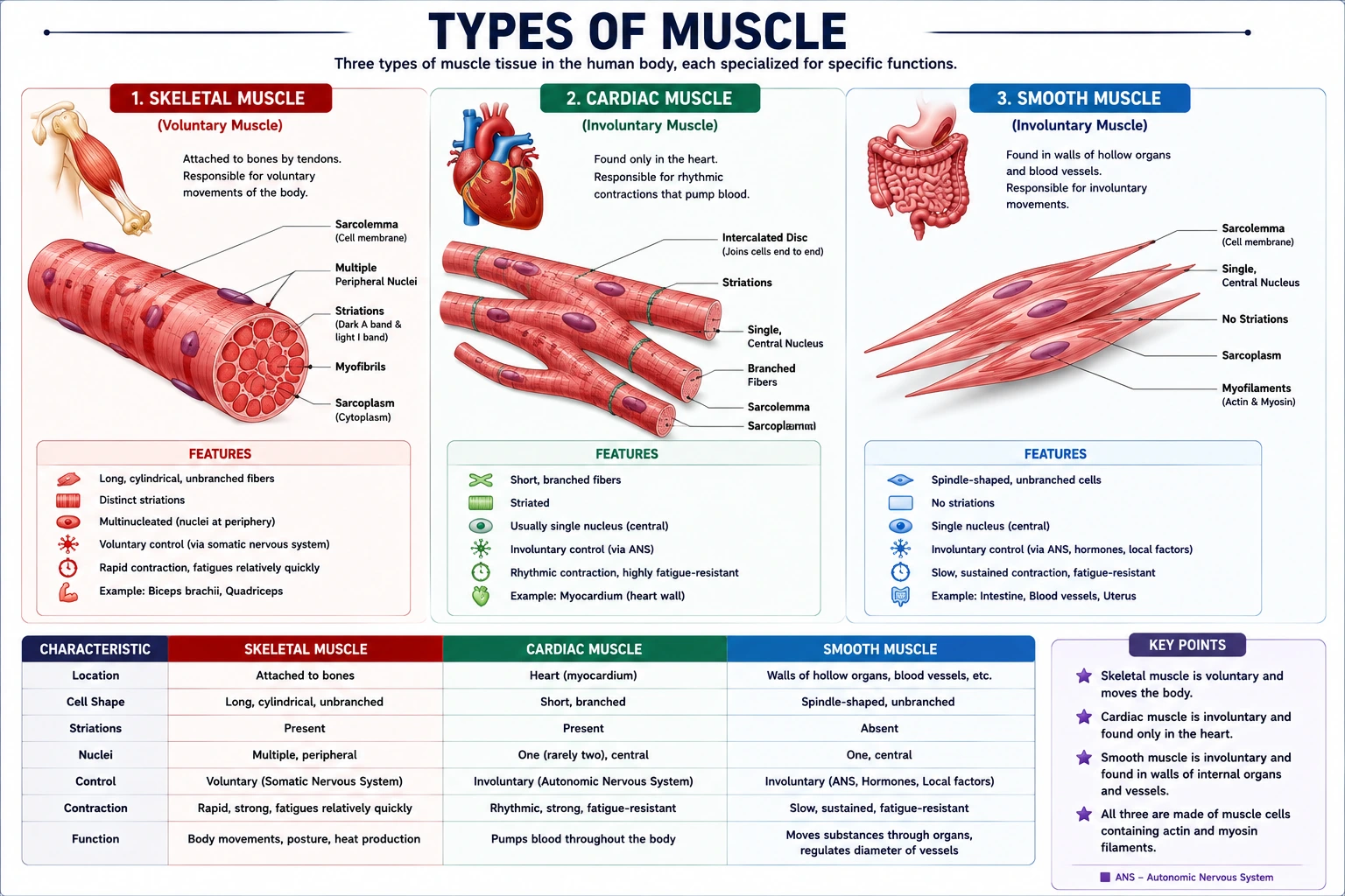
The human body contains three distinct types of muscle tissue, each specialized for particular functions:
- Skeletal Muscle – Voluntary, striated muscle attached to bones; responsible for locomotion, posture, and heat generation.
- Smooth Muscle – Involuntary, non‑striated muscle found in the walls of hollow organs (blood vessels, gastrointestinal tract, bladder, uterus); controls internal movements such as peristalsis and vasoconstriction.
- Cardiac Muscle – Involuntary, striated muscle found exclusively in the heart; responsible for pumping blood throughout the body.
Despite their differences, all muscle types share the fundamental property of contractility – the ability to shorten and generate force. The following table summarizes their key characteristics.
Comparison of Muscle Tissue Types
| Feature | Skeletal Muscle | Smooth Muscle | Cardiac Muscle |
|---|---|---|---|
| Location | Attached to bones | Walls of hollow organs (gut, vessels, bladder, uterus) | Heart only |
| Control | Voluntary | Involuntary | Involuntary |
| Striations | Yes (highly organized sarcomeres) | No (actin and myosin not arranged in sarcomeres) | Yes (striated, but less regular than skeletal) |
| Nucleus | Multinucleated (peripheral) | Single, centrally located | Single (or occasionally binucleated), centrally located |
| T-tubules | Well developed, at A‑I junction | Absent (caveolae instead) | Well developed, at Z‑disc, larger diameter |
| Sarcoplasmic Reticulum | Extensive, well organized | Poorly developed; Ca²⁺ enters mainly from extracellular fluid | Moderate; Ca²⁺ from both SR and extracellular space |
| Source of Ca²⁺ for contraction | Sarcoplasmic reticulum (SR) only | Extracellular fluid and SR (mainly extracellular) | SR and extracellular fluid |
| Regulation of Contraction | Troponin‑tropomyosin system | Calmodulin‑myosin light chain kinase (MLCK) system | Troponin‑tropomyosin system (similar to skeletal) |
| Gap Junctions | No (each fiber stimulated independently) | Yes (single‑unit smooth muscle) – allows coordinated contraction | Yes (intercalated discs) – functional syncytium |
| Autorhythmicity | No (requires nerve stimulation) | Yes (some types, e.g., pacemaker cells in gut) | Yes (pacemaker cells in SA node) |
| Speed of Contraction | Fast to slow (depending on fiber type) | Very slow | Moderate (slower than skeletal, faster than smooth) |
| Energy Source | ATP (oxidative and glycolytic) | ATP (mainly glycolytic) | ATP (mainly oxidative, rich in mitochondria) |
| Regeneration Ability | Limited (satellite cells) | High (mitosis of smooth muscle cells) | Very limited (no mitosis in adults) |
Core Content
1. Physiological Anatomy of Skeletal Muscle
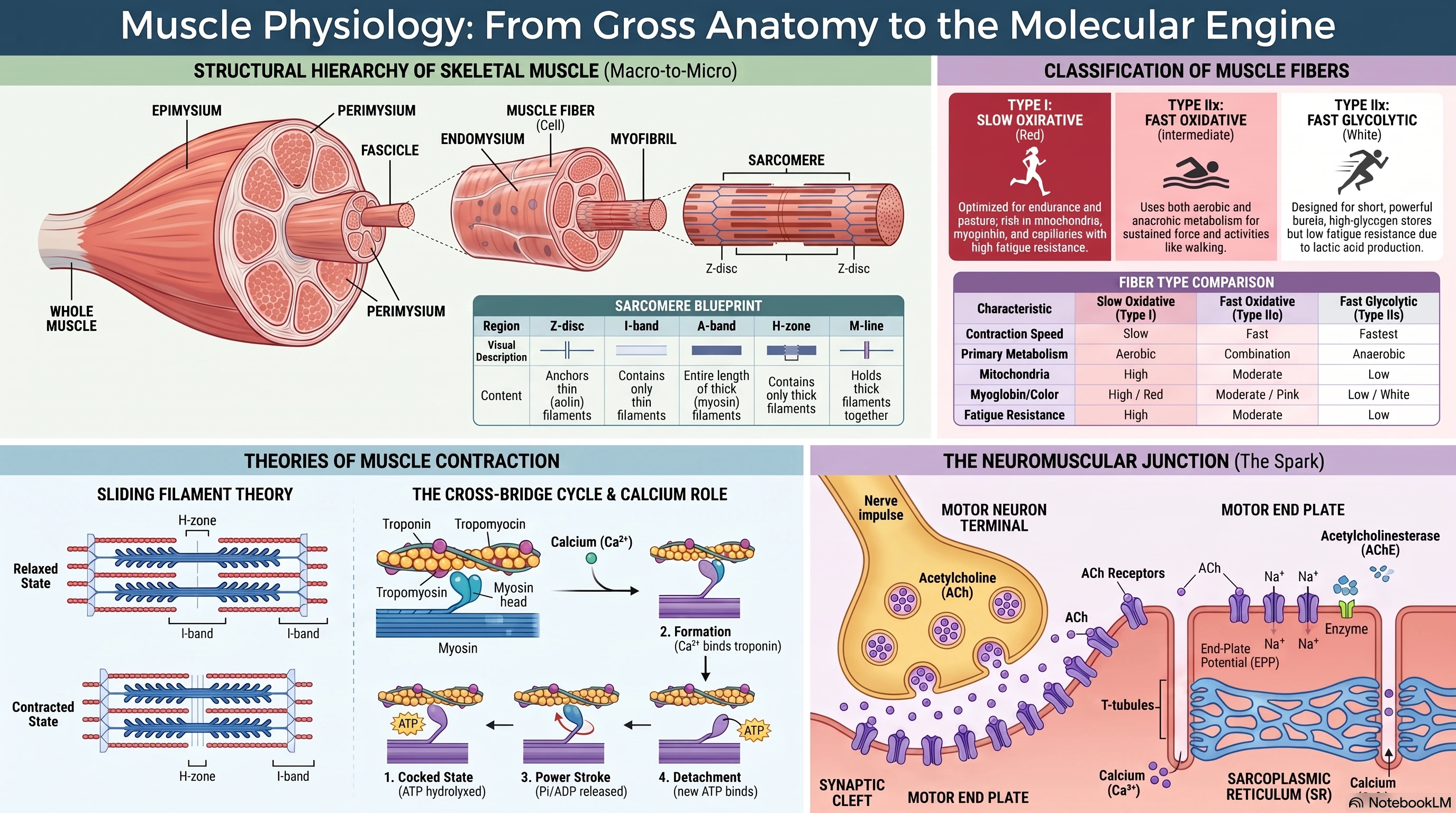
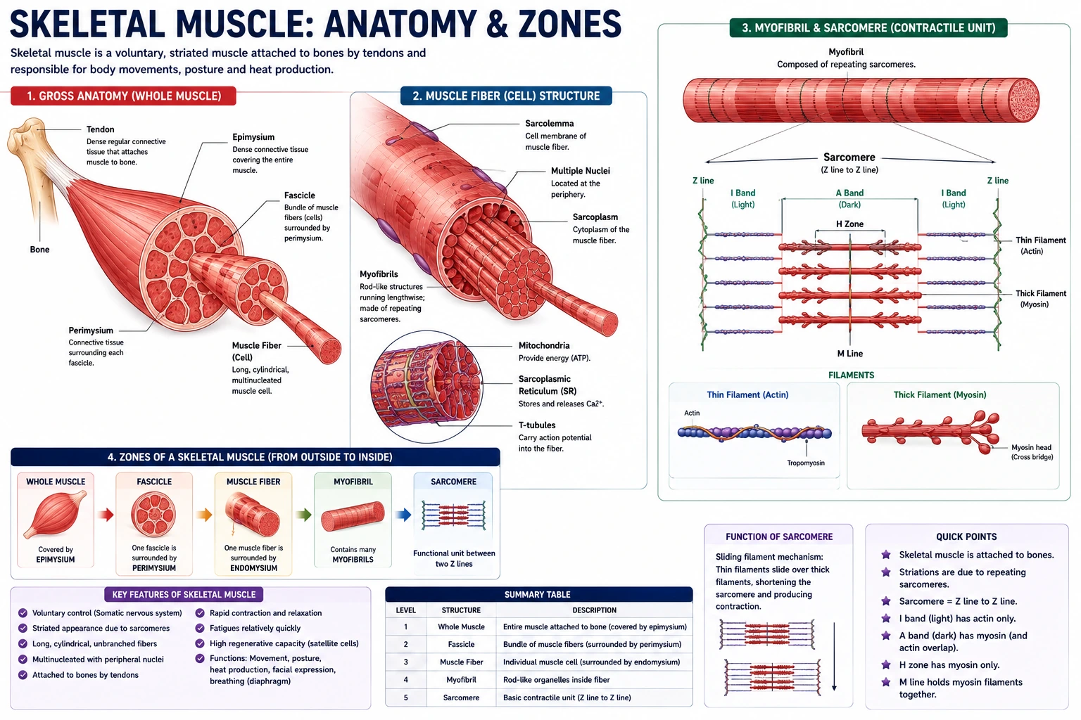
Skeletal muscle is a highly organized tissue responsible for voluntary movement, posture, and heat production. Its structure is hierarchical, enabling efficient force generation.
A. Gross Anatomy (Organ Level)
Each skeletal muscle is an organ composed of muscle tissue, blood vessels, nerve fibers, and connective tissues. The connective tissue layers organize and support the muscle:
- Epimysium: The outermost layer that surrounds the entire muscle organ. It allows the muscle to contract and move powerfully while maintaining structural integrity.
- Perimysium: The connective tissue that separates muscle fibers into bundles called fascicles. This organization allows the nervous system to activate specific subsets of fibers for precise movements.
- Endomysium: A thin layer of connective tissue encasing each individual muscle fiber. It contains extracellular fluid and supplies nutrients.
These connective tissue layers are continuous with tendons, which attach muscles to bones.
B. Microscopic Anatomy (Muscle Fiber/Cell Level)
A skeletal muscle fiber is a single, multinucleated cell formed by the fusion of hundreds of myoblasts during development. It has specialized terminology:
- Sarcolemma: The plasma membrane of the muscle fiber.
- Sarcoplasm: The cytoplasm of the muscle fiber.
- Sarcoplasmic Reticulum (SR): A specialized smooth endoplasmic reticulum that stores and releases calcium ions (Ca²⁺), essential for contraction.
- Transverse (T) Tubules: Invaginations of the sarcolemma that extend deep into the muscle fiber. They transmit action potentials from the cell surface to the interior.
Within the sarcoplasm are myofibrils, the contractile organelles. Each myofibril is a thread‑like structure composed of repeating units called sarcomeres.
C. The Sarcomere (Functional Unit)
The sarcomere is the basic contractile unit of skeletal muscle, extending from one Z‑disc to the next. It contains two types of protein filaments:
- Thick Filaments: Composed of the protein myosin.
- Thin Filaments: Composed primarily of actin.
The arrangement of these filaments creates the striated pattern of skeletal muscle. Key regions of the sarcomere include:
| Region | Description |
|---|---|
| Z‑disc (Z‑line) | Boundary of the sarcomere; anchors thin filaments. |
| I‑band | Light‑colored region containing only thin filaments; bisected by the Z‑disc. |
| A‑band | Dark‑colored region containing the entire length of thick filaments (and overlapping thin filaments). |
| H‑zone | Lighter region in the center of the A‑band containing only thick filaments. |
| M‑line | The center of the sarcomere; holds thick filaments together. |
2. Types of Skeletal Muscle Fibers
Skeletal muscle fibers are classified based on two main criteria: their speed of contraction and their primary metabolic pathway for generating ATP.
A. Slow Oxidative (Type I, Slow‑Twitch)
- Contraction Speed: Slow.
- Metabolism: Aerobic (oxidative). Rich in mitochondria and myoglobin, giving them a red color. Extensively supplied with capillaries.
- Fatigue Resistance: High. Can contract for long periods without fatiguing.
- Function: Maintain posture, stabilize bones and joints. Used for endurance activities.
B. Fast Oxidative (Type IIa, Fast‑Twitch Oxidative‑Glycolytic)
- Contraction Speed: Fast.
- Metabolism: Combination of aerobic and anaerobic. Intermediate numbers of mitochondria and moderate myoglobin content.
- Fatigue Resistance: Moderate to high.
- Function: Used for movements requiring more energy than postural control, such as walking.
C. Fast Glycolytic (Type IIx, Fast‑Twitch Anaerobic)
- Contraction Speed: Fastest.
- Metabolism: Anaerobic (glycolytic). Few mitochondria, low myoglobin (white color), large glycogen stores. Limited capillary supply.
- Fatigue Resistance: Low. Fatigue quickly due to lactic acid production and inefficient ATP production.
- Function: Produces rapid, forceful contractions for short, powerful movements.
Most human muscles contain a mixture of all three fiber types.
3. General and Molecular Mechanisms of Muscle Contraction
A. The Sliding Filament Theory
Muscle contraction occurs when sarcomeres shorten. According to the sliding filament theory, the thick and thin filaments do not change length; instead, they slide past each other, increasing the degree of overlap. This causes the Z‑discs to move closer together, shortening the sarcomere and the entire muscle fiber. The I‑bands and H‑zones shorten, while the A‑bands remain the same length.
B. The Cross‑Bridge Cycle (Molecular Mechanism)
The sliding of filaments is driven by a cyclical interaction between myosin heads and actin filaments, powered by ATP.
- Cocked State: A myosin head, with bound ATP hydrolyzed to ADP and Pi (inorganic phosphate), is in a high‑energy, “cocked” conformation. The myosin‑binding sites on actin are blocked by the tropomyosin‑troponin complex.
- Cross‑Bridge Formation: When calcium ions (Ca²⁺) bind to troponin, it causes a conformational change that moves tropomyosin away from the myosin‑binding sites on actin. The energized myosin head binds to the exposed site, forming a cross‑bridge.
- Power Stroke: The release of Pi and ADP triggers the myosin head to pivot, pulling the actin filament toward the center of the sarcomere. This is the “power stroke” that generates force.
- Detachment: A new ATP molecule binds to the myosin head, causing it to detach from the actin filament.
- Re‑cocking: ATP is hydrolyzed, providing the energy to re‑cock the myosin head, returning it to its high‑energy state and readying it for another cycle.
This cycle repeats as long as Ca²⁺ is available and ATP is present.
C. Excitation‑Contraction Coupling
This process links the electrical signal from the nervous system to the mechanical contraction of the muscle.
- Nerve Stimulus: An action potential travels down a motor neuron to the neuromuscular junction.
- Neuromuscular Transmission: The action potential triggers the release of the neurotransmitter acetylcholine (ACh) into the synaptic cleft. ACh binds to receptors on the motor end plate of the muscle fiber, generating an end‑plate potential.
- Muscle Action Potential: The end‑plate potential triggers an action potential in the muscle fiber’s sarcolemma.
- Signal Propagation: The action potential propagates along the sarcolemma and down into the T‑tubules.
- Calcium Release: The action potential in the T‑tubules triggers the sarcoplasmic reticulum (SR) to release large amounts of stored Ca²⁺ into the sarcoplasm.
- Contraction: The released Ca²⁺ binds to troponin, initiating the cross‑bridge cycle and muscle contraction.
- Relaxation: When stimulation stops, the SR pumps Ca²⁺ back into its stores using active transport (ATP‑dependent). The drop in intracellular Ca²⁺ causes tropomyosin to re‑cover the myosin‑binding sites, and the muscle relaxes.
4. Physiological Anatomy of the Neuromuscular Junction
The neuromuscular junction (NMJ) is the specialized synapse between a somatic motor neuron and a skeletal muscle fiber. It is the only point of neural control for skeletal muscle contraction.
Key Structures:
- Motor Neuron Terminal (Presynaptic): The enlarged, unmyelinated ending of the motor neuron. It contains synaptic vesicles filled with the neurotransmitter acetylcholine (ACh).
- Synaptic Cleft: The narrow gap (approximately 50 nanometers) between the neuron and the muscle fiber.
- Motor End Plate (Postsynaptic): The highly folded region of the sarcolemma directly across from the neuron. It is densely packed with ACh receptors.
- Acetylcholinesterase (AChE): An enzyme located on the postsynaptic membrane that rapidly breaks down ACh, ensuring a single nerve impulse produces a single muscle contraction.
Process of Neuromuscular Transmission:
- An action potential reaches the motor neuron terminal.
- The depolarization opens voltage‑gated calcium (Ca²⁺) channels in the presynaptic membrane. Ca²⁺ enters the terminal.
- The influx of Ca²⁺ triggers the exocytosis of synaptic vesicles, releasing ACh into the synaptic cleft.
- ACh diffuses across the cleft and binds to ACh receptors on the motor end plate.
- These receptors are ligand‑gated cation channels. Their opening allows Na⁺ to enter the muscle fiber and K⁺ to leave. The net influx of Na⁺ creates a depolarization called the end‑plate potential (EPP).
- The EPP is always suprathreshold, meaning it always triggers an action potential in the adjacent sarcolemma.
- The action potential then propagates across the muscle fiber surface and into the T‑tubules, initiating excitation‑contraction coupling.
- ACh is rapidly degraded by AChE, terminating the signal and allowing the muscle fiber to repolarize.
5. The Action Potential in Skeletal Muscle
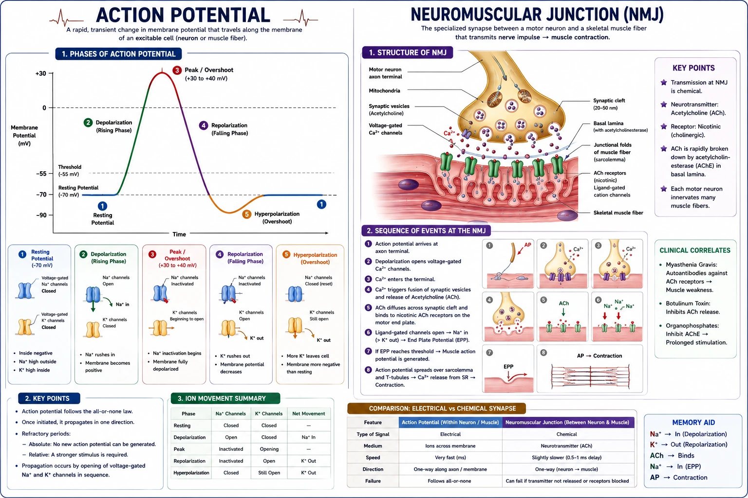
The action potential is the electrical signal that triggers muscle contraction. It is a rapid, transient change in the membrane potential that propagates along the sarcolemma and into the T‑tubules, ensuring the entire fiber contracts synchronously.
A. Resting Membrane Potential (RMP)
The inside of a resting skeletal muscle fiber is about ‑80 to ‑90 mV relative to the outside. This potential is maintained by:
- Na⁺/K⁺ ATPase pump – actively transports 3 Na⁺ out and 2 K⁺ in per ATP hydrolyzed.
- Differential permeability – at rest, the membrane is far more permeable to K⁺ than Na⁺, allowing K⁺ to leak out, making the interior negative.
- Fixed anions – negatively charged proteins and organic phosphates inside the cell.
B. Phases of the Skeletal Muscle Action Potential
| Phase | Ionic Event | Membrane Potential |
|---|---|---|
| Resting | K⁺ efflux through leak channels; Na⁺/K⁺ pump maintains gradients. | –90 mV |
| Threshold | Sufficient depolarization (e.g., from end‑plate potential) opens voltage‑gated Na⁺ channels. | ~ –65 mV |
| Depolarization | Rapid Na⁺ influx through voltage‑gated Na⁺ channels → membrane potential shoots up to +30 mV. | –90 to +30 mV |
| Repolarization | Voltage‑gated Na⁺ channels inactivate; voltage‑gated K⁺ channels open → K⁺ efflux restores negative interior. | +30 mV back toward –90 mV |
| After‑hyperpolarization | K⁺ channels close slowly, causing a brief undershoot below RMP. | ~ –95 mV |
| Return to RMP | Na⁺/K⁺ pump and leak channels restore resting state. | –90 mV |
Key Points:
- The action potential in skeletal muscle is very similar to that in neurons, but lasts longer (~2‑5 ms).
- The depolarization phase is due to voltage‑gated Na⁺ channels opening, while repolarization is due to voltage‑gated K⁺ channels opening and Na⁺ channel inactivation.
- The action potential is all‑or‑none – once threshold is reached, a full action potential is generated.
- After an action potential, there is a brief absolute refractory period (no stimulus can trigger another AP) followed by a relative refractory period (a stronger‑than‑normal stimulus is required).
C. Propagation of the Action Potential
- The action potential is generated at the motor end plate (NMJ).
- It spreads bidirectionally along the sarcolemma.
- The depolarization wave enters the T‑tubules, which carry it deep into the fiber.
- At the T‑tubules, dihydropyridine (DHP) receptors (voltage sensors) trigger Ca²⁺ release from the sarcoplasmic reticulum via ryanodine receptors, linking the action potential to contraction (excitation‑contraction coupling).
D. Comparison: Action Potential in Nerve vs. Skeletal Muscle
| Feature | Nerve (Axon) | Skeletal Muscle |
|---|---|---|
| Resting potential | –70 mV | –80 to –90 mV |
| Duration | ~1 ms | ~2‑5 ms |
| Main depolarizing ion | Na⁺ (voltage‑gated) | Na⁺ (voltage‑gated) |
| Repolarizing ion | K⁺ (voltage‑gated) | K⁺ (voltage‑gated) |
| Propagation speed | Up to 120 m/s (myelinated) | ~3‑5 m/s (along sarcolemma) |
| Role of Ca²⁺ | Triggers neurotransmitter release | Triggers contraction |
Tables
Table 1: Comparison of Skeletal Muscle Fiber Types
| Characteristic | Slow Oxidative (Type I) | Fast Oxidative (Type IIa) | Fast Glycolytic (Type IIx) |
|---|---|---|---|
| Other Names | Slow‑Twitch, Red | Fast‑Twitch, Intermediate | Fast‑Twitch, White |
| Contraction Speed | Slow | Fast | Fastest |
| Primary Metabolism | Aerobic (Oxidative) | Combination (Aerobic/Anaerobic) | Anaerobic (Glycolytic) |
| ATPase Activity | Low | High | Highest |
| Mitochondria | High | Moderate | Low |
| Myoglobin Content | High | Moderate | Low |
| Glycogen Content | Low | Moderate | High |
| Capillary Supply | Rich | Moderate | Poor |
| Fatigue Resistance | High | Moderate | Low |
| Function | Posture, Endurance | Walking, Sustained force | Powerful, Short bursts |
Table 2: Steps of the Cross‑Bridge Cycle
| Step | Event | Key Molecules |
|---|---|---|
| 1. Cocked State | Myosin head is energized (ADP + Pi bound). Binding sites on actin are blocked by tropomyosin. | Myosin, Actin, ADP, Pi, Tropomyosin, Troponin |
| 2. Cross‑Bridge Formation | Ca²⁺ binds to troponin, moving tropomyosin. Myosin head binds to exposed site on actin. | Myosin, Actin, Ca²⁺, Troponin |
| 3. Power Stroke | Myosin head pivots, pulling actin filament. Pi and ADP are released. | Myosin, Actin |
| 4. Detachment | ATP binds to myosin head, causing it to detach from actin. | Myosin, Actin, ATP |
| 5. Re‑cocking | ATP is hydrolyzed to ADP + Pi, energy is stored in the myosin head. | Myosin, ATP → ADP + Pi |
Exam Angle
Short Answer Questions
- Define a sarcomere and draw and label its major regions (Z‑disc, I‑band, A‑band, H‑zone, M‑line).
- Differentiate between the three types of skeletal muscle fibers based on their structure and function.
- Describe the structure of the neuromuscular junction.
- What is the role of calcium ions in muscle contraction?
- Explain the sliding filament theory of muscle contraction.
- Describe the phases of the action potential in skeletal muscle.
Essay Questions
- Describe the molecular mechanisms of muscle contraction, detailing the cross‑bridge cycle, excitation‑contraction coupling, and the role of ATP.
- Trace the sequence of events from an action potential arriving at the neuromuscular junction to the relaxation of a skeletal muscle fiber.
- Compare and contrast the three types of skeletal muscle fibers.
Viva / Short Notes
- Excitation‑Contraction Coupling.
- Structure and function of the sarcoplasmic reticulum.
- The role of troponin and tropomyosin.
- The neuromuscular junction.
- Motor unit.
- Action potential propagation in T‑tubules.
Summary Box
- Skeletal muscle is organized hierarchically: whole muscle → fascicles → muscle fibers (cells) → myofibrils → sarcomeres. The sarcomere is the functional unit of contraction, containing interdigitating actin (thin) and myosin (thick) filaments.
- Skeletal muscle fibers are classified into Type I (slow oxidative) for endurance, Type IIa (fast oxidative) for intermediate activities, and Type IIx (fast glycolytic) for powerful, short‑duration movements.
- The sliding filament theory describes contraction as the sliding of actin filaments past myosin filaments, shortening the sarcomere without changing filament length.
- The cross‑bridge cycle, driven by ATP, involves myosin heads binding to actin, performing a power stroke, detaching, and re‑cocking. This is regulated by Ca²⁺ binding to troponin, which uncovers the myosin‑binding sites on actin.
- Excitation‑contraction coupling connects the neural signal to contraction: ACh released at the neuromuscular junction triggers a muscle action potential that travels down T‑tubules, causing the SR to release Ca²⁺.
- The neuromuscular junction is a highly specialized chemical synapse where a motor neuron communicates with a muscle fiber via acetylcholine (ACh). The signal is terminated by acetylcholinesterase to allow relaxation.
- The action potential of skeletal muscle consists of a rapid depolarization (Na⁺ influx) followed by repolarization (K⁺ efflux); it propagates along the sarcolemma and into T‑tubules to trigger Ca²⁺ release.
References
- Hall, J. E., & Hall, M. E. (2021). Guyton and Hall Textbook of Medical Physiology (14th ed.). Elsevier.
- Barrett, K. E., Barman, S. M., Boitano, S., & Brooks, H. L. (2019). Ganong’s Review of Medical Physiology (26th ed.). McGraw‑Hill Education.
- Boron, W. F., & Boulpaep, E. L. (2017). Medical Physiology (3rd ed.). Elsevier.
- Sherwood, L. (2016). Human Physiology: From Cells to Systems (9th ed.). Cengage Learning.
- Costanzo, L. S. (2018). Physiology (6th ed.). Elsevier.
End of unit 2 of 7
Check what you've learned
5 quick questions from this unit. They're optional: try them if you like, then mark the unit as read and move on.