Unit 4: The Heart
Cardiac cycle, ECG, regulation of heart function.
Prepared by the PharmaWallah student team. Verify results with your textbook or instructor. Report an error · Editorial policy
Unit 4: The Heart
Learning Objectives
- Describe the physiological anatomy of heart muscle and the structure of the heart.
- Trace the course of blood flow through the heart and explain the cardiac cycle.
- Describe the conduction system of the heart and its role in generating rhythmic contractions.
- Explain the production of heart sounds and murmurs and their clinical significance.
- Interpret the characteristics of a normal electrocardiogram (ECG) and its relationship to the cardiac cycle.
- Define and apply key hemodynamic terms: systole, diastole, stroke volume, cardiac output, preload, afterload, and ejection fraction.
Core Content
1. Physiological Anatomy of Heart Muscle
The heart is a dual pump composed of specialised cardiac muscle (myocardium). It lies in the mediastinum, enclosed by the pericardium.
A. Characteristics of Cardiac Muscle
| Feature | Cardiac Muscle | Skeletal Muscle | Smooth Muscle |
|---|---|---|---|
| Striations | Yes | Yes | No |
| Nuclei | Single, central | Multiple, peripheral | Single, central |
| Cell shape | Branched, cylindrical | Long, cylindrical | Spindle‑shaped |
| Intercalated discs | Yes (gap junctions + desmosomes) | No | No (gap junctions in some) |
| Automaticity | Yes (pacemaker cells) | No | Yes (some) |
| Action potential | Long (200–400 ms) with plateau | Short (2–5 ms) | Variable |
| Tetanus | Cannot occur (long refractory) | Can occur | Can occur |
| Control | Involuntary, intrinsic | Voluntary | Involuntary |
Key specialisations:
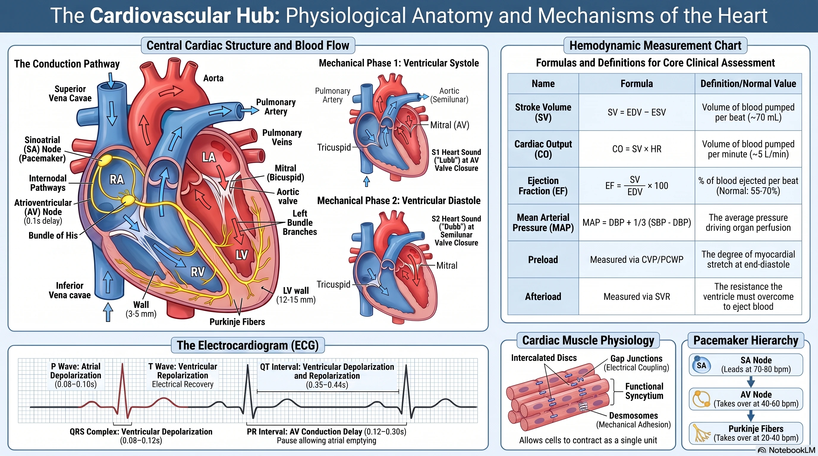
- Intercalated discs contain gap junctions (electrical coupling) and desmosomes (mechanical adhesion).
- The heart works as two functional syncytia – atrial and ventricular – separated by the fibrous skeleton. The only electrical connection is the AV bundle.
B. Cardiac Muscle Action Potential
The ventricular action potential has a prolonged plateau (Phase 2) due to slow Ca²⁺ channels, preventing tetanus and allowing complete filling.
| Phase | Name | Ion movement |
|---|---|---|
| 4 | Resting membrane potential | Stable at –90 mV; K⁺ efflux via I<sub>K1</sub> channels |
| 0 | Rapid depolarisation | Voltage‑gated Na⁺ channels open → Na⁺ influx |
| 1 | Early repolarisation | Transient K⁺ efflux (I<sub>to</sub>) |
| 2 | Plateau | L‑type Ca²⁺ channels open (I<sub>Ca,L</sub>); K⁺ efflux delayed |
| 3 | Repolarisation | Ca²⁺ channels inactivate; delayed rectifier K⁺ channels open |
Refractory periods:
- Absolute refractory period – no new action potential possible.
- Effective refractory period – only a local response possible.
- Relative refractory period – stronger‑than‑normal stimulus required.
Pacemaker action potential (SA node):
- Unstable resting potential, spontaneous diastolic depolarisation due to funny current (I<sub>f</sub>) and T‑type Ca²⁺ channels.
- Upstroke via L‑type Ca²⁺ channels (not Na⁺).
| Cell type | Resting potential | Upstroke | Plateau | Intrinsic rate (min⁻¹) |
|---|---|---|---|---|
| SA node | –60 mV (unstable) | Slow (Ca²⁺) | No | 60–100 |
| Atrial muscle | –80 mV | Fast (Na⁺) | Short | Follows SA node |
| AV node | –60 mV (unstable) | Slow (Ca²⁺) | No | 40–60 (conduction delay) |
| Purkinje fibres | –90 mV | Fast (Na⁺) | Yes | 20–40 (fast conduction) |
| Ventricular muscle | –90 mV | Fast (Na⁺) | Yes | Follows Purkinje |
C. Excitation‑Contraction Coupling (Cardiac)
- Action potential → L‑type Ca²⁺ channels open → trigger Ca²⁺ influx → calcium‑induced calcium release (CICR) from SR via ryanodine receptors (RyR2).
- Relaxation: Ca²⁺ re‑uptake into SR (SERCA2a) and extrusion via Na⁺/Ca²⁺ exchanger (NCX).
- Force‑frequency relationship: Higher heart rate → more Ca²⁺ influx → increased contractility (Bowditch effect).
Pharmacy Note: Drugs that alter Ca²⁺ handling (e.g., β‑blockers, calcium channel blockers, digoxin) directly affect cardiac contractility and rate.
2. Structure of the Heart
A. Chambers and Wall Thickness
| Chamber | Function | Wall thickness |
|---|---|---|
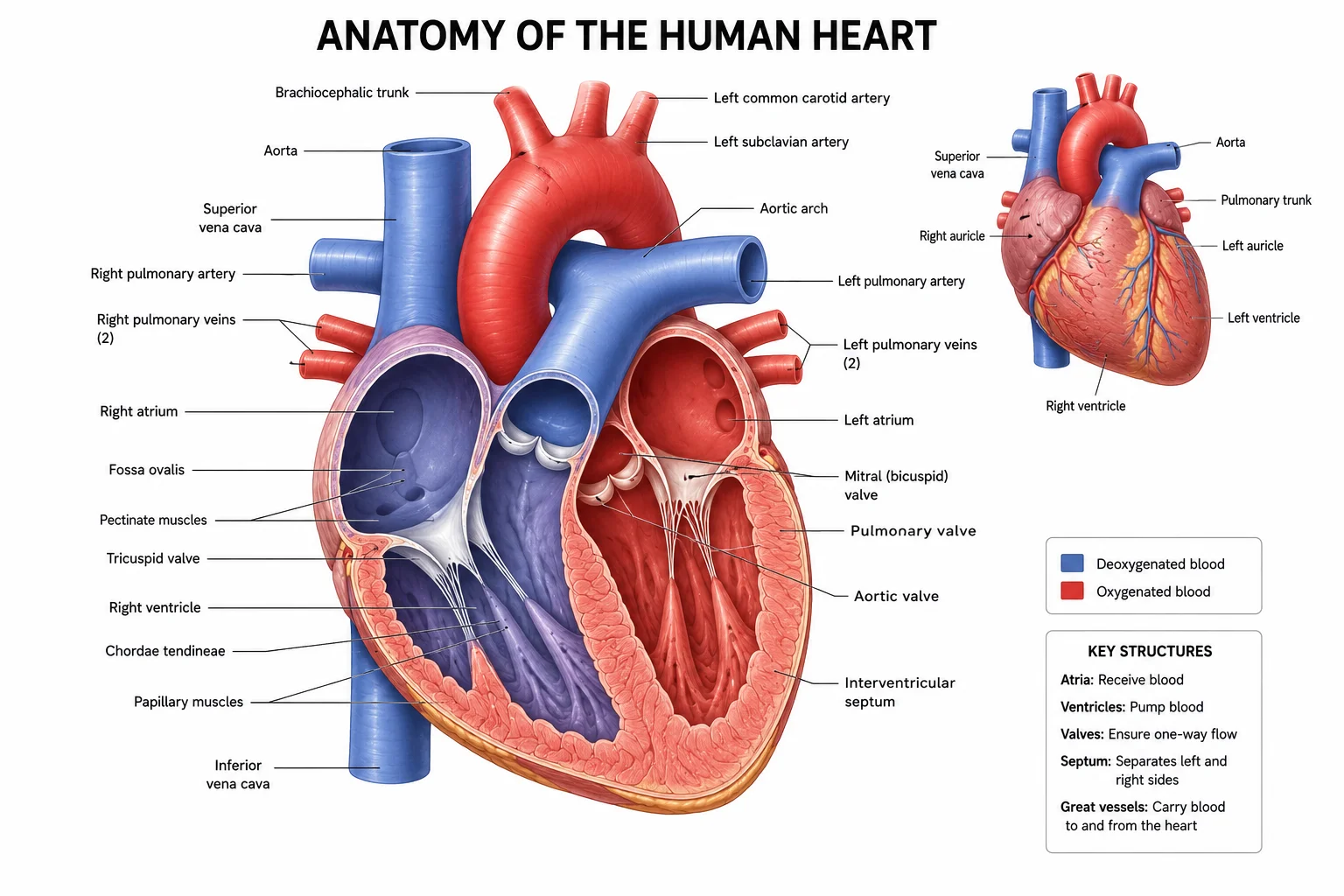
| Right atrium | Receives deoxygenated blood (SVC, IVC, coronary sinus) | Thin |
| Right ventricle | Pumps blood to lungs (pulmonary circulation) | Moderate (3–5 mm) |
| Left atrium | Receives oxygenated blood (pulmonary veins) | Thin |
| Left ventricle | Pumps blood to body (systemic circulation) | Thick (8–15 mm) |
B. Heart Valves
| Valve | Location | Type | Prevents backflow during |
|---|---|---|---|
| Tricuspid | Right atrium → right ventricle | AV valve | Ventricular systole |
| Pulmonary (semilunar) | Right ventricle → pulmonary trunk | Semilunar valve | Ventricular diastole |
| Mitral (bicuspid) | Left atrium → left ventricle | AV valve | Ventricular systole |
| Aortic (semilunar) | Left ventricle → aorta | Semilunar valve | Ventricular diastole |
Valve pathology:
- Stenosis – narrowed opening, impedes forward flow.
- Regurgitation (insufficiency) – incomplete closure, allows backward flow.
C. Coronary Circulation
| Artery | Branches | Territory supplied |
|---|---|---|
| Left main coronary | LAD, LCx | |
| LAD | Septal perforators, diagonals | Anterior septum, anterior LV |
| LCx | Obtuse marginal | Lateral/posterior LV |
| Right coronary (RCA) | Acute marginal, PDA (85% of people) | RV, SA node (60%), AV node (80%), inferior LV |
Coronary blood flow:
- ~250 mL/min at rest (5% of CO), increases 4–5× with exercise.
- Flow mainly during diastole (ventricular compression of vessels during systole).
- Regulated metabolically (adenosine, NO, O₂, CO₂).
3. Course of Blood Flow Through the Heart
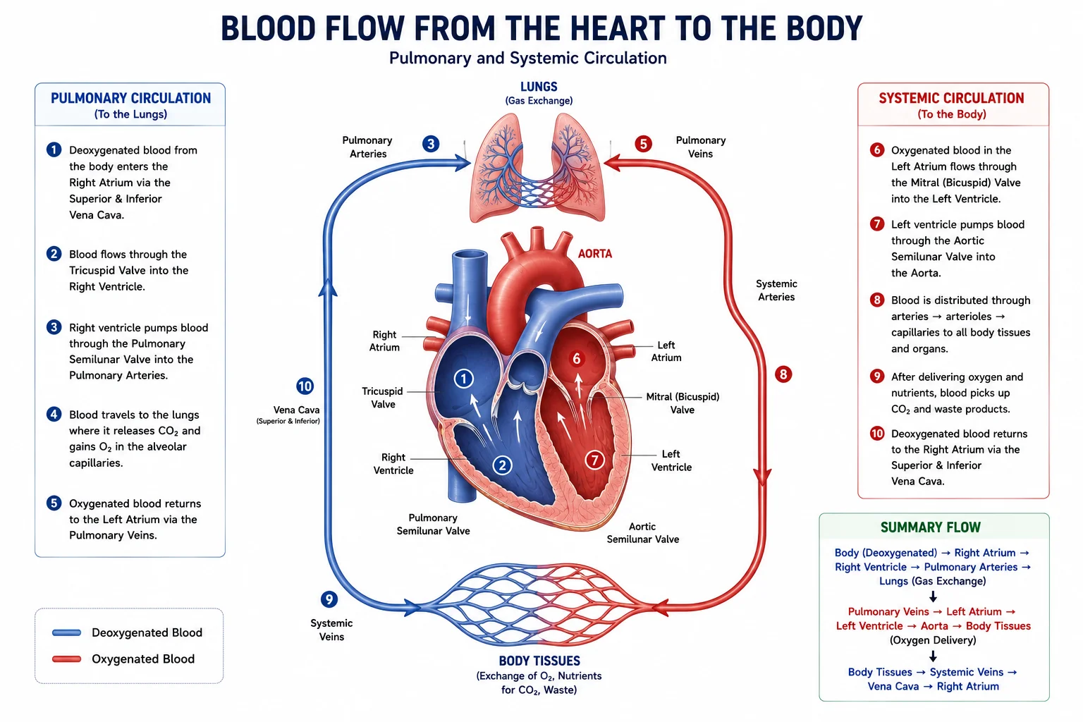
- Right side handles deoxygenated blood (pulmonary circulation).
- Left side handles oxygenated blood (systemic circulation). Left ventricular wall is thicker because it pumps against higher pressure.
4. The Cardiac Cycle
At a heart rate of 75 bpm, cycle length ≈ 0.8 s (systole ~0.3 s, diastole ~0.5 s).
| Phase | AV valves | Semilunar valves | Ventricular pressure | Ventricular volume | ECG |
|---|---|---|---|---|---|
| Ventricular systole | |||||
| 1. Isovolumetric contraction | Closed | Closed | ↑↑ rapidly | Constant (EDV) | After QRS |
| 2. Rapid ejection | Closed | Open | Peaks | ↓↓ rapidly | ST segment |
| 3. Reduced ejection | Closed | Open | ↓ slightly | ↓ slowly | End of T |
| Ventricular diastole | |||||
| 4. Isovolumetric relaxation | Closed | Closed | ↓↓ rapidly | Constant (ESV) | End of T |
| 5. Rapid filling | Open | Closed | Low | ↑↑ rapidly | After T |
| 6. Reduced filling (diastasis) | Open | Closed | Low | ↑ slowly | Before P |
| 7. Atrial systole | Open | Closed | Slight ↑ | ↑ to EDV | After P |
Pressure‑volume loop (not drawn):
- A (mitral valve closes): EDV ~120 mL.
- B (aortic valve opens): diastole ends.
- C (aortic valve closes): ESV ~50 mL.
- D (mitral valve opens): ESV, filling begins.
5. Conduction System of the Heart
| Component | Location | Function | Intrinsic rate (min⁻¹) |
|---|---|---|---|
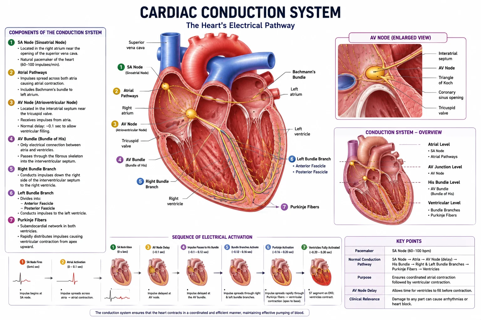
| SA node | Right atrium near SVC | Primary pacemaker; initiates impulse | 60–100 |
| Internodal pathways | Atrial wall | Conduct impulse to AV node | – |
| AV node | Interatrial septum | Delays impulse (~0.1 s) | 40–60 |
| Bundle of His | Interventricular septum | Sole electrical bridge between atria/ventricles | – |
| Bundle branches | Septum | Conduct to Purkinje | – |
| Purkinje fibres | Subendocardial of ventricles | Rapid depolarisation of ventricles | 20–40 |
Conduction velocities:
- Atrial muscle: 0.3–0.5 m/s
- AV node: 0.05 m/s (slowest – ensures delay)
- Purkinje fibres: 2–4 m/s (fastest)
Autonomic regulation:
- Sympathetic (β₁) → ↑ HR (positive chronotropy), ↑ conduction, ↑ contractility (via cAMP, I<sub>f</sub>, Ca²⁺).
- Parasympathetic (vagus, M₂) → ↓ HR, ↓ AV conduction (via ↓ cAMP, ↑ K⁺ conductance).
Pharmacy Note: β‑blockers, calcium channel blockers (verapamil/diltiazem), and digoxin are commonly used to control heart rate in arrhythmias.
6. Heart Sounds and Murmurs
A. Normal Heart Sounds
| Sound | Cause | Timing | Auscultation |
|---|---|---|---|
| S₁ | Closure of AV valves (mitral & tricuspid) | Beginning of systole | Apex (mitral), lower left sternal border (tricuspid) |
| S₂ | Closure of semilunar valves (aortic & pulmonary) | Beginning of diastole | 2nd ICS right (aortic), 2nd ICS left (pulmonary) |
| S₃ | Rapid ventricular filling | Early diastole | Normal in children/youth; pathological in HF |
| S₄ | Atrial systole against stiff ventricle | Late diastole (before S₁) | Pathological (hypertension, AS) |
Splitting of S₂:
- Physiological – during inspiration (increased RV ejection time).
- Paradoxical – during expiration (LV ejection delayed; e.g., LBBB).
- Fixed – atrial septal defect (no respiratory change).
B. Heart Murmurs (Turbulent flow)
| Timing | Common causes |
|---|---|
| Systolic | Aortic stenosis (ejection, crescendo‑decrescendo), mitral regurgitation (holosystolic), VSD |
| Diastolic | Aortic regurgitation (early decrescendo), mitral stenosis (mid‑diastolic rumble) |
| Continuous | Patent ductus arteriosus (machinery murmur) |
Grading (1–6):
- Barely audible
- Soft but readily heard
- Moderately loud
- Loud + thrill
- Very loud (stethoscope edge)
- Heard without stethoscope
7. Electrocardiogram (ECG)
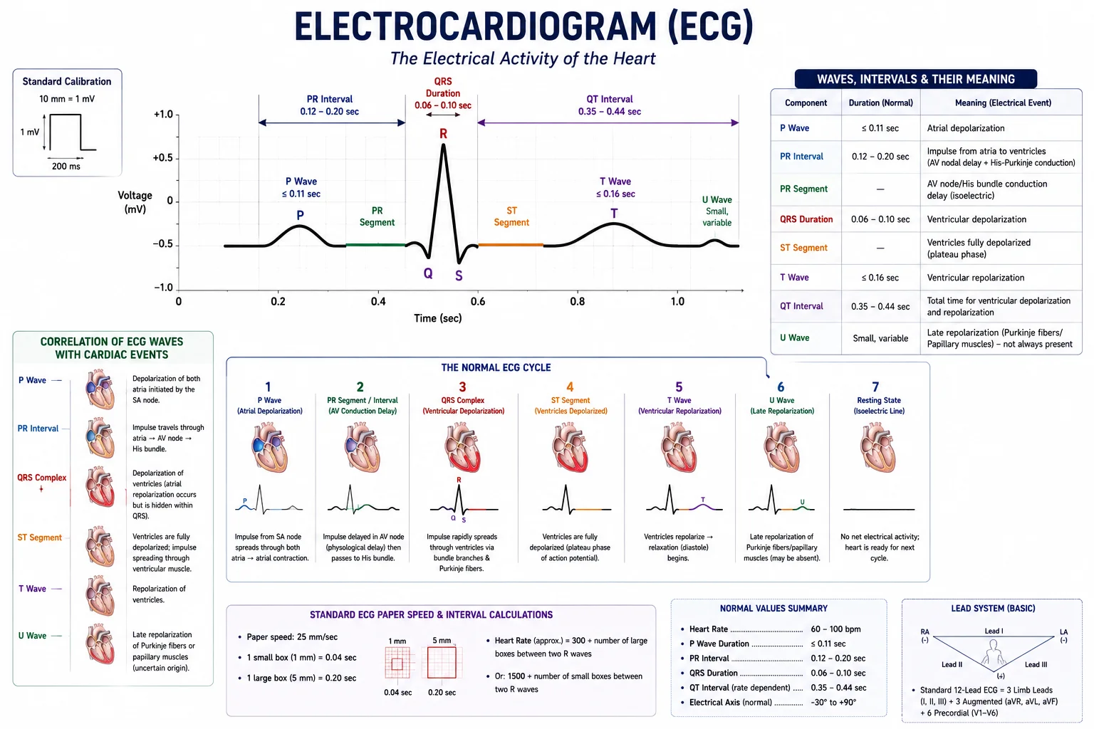
A. Normal ECG Waves
| Wave/Interval | Duration | Meaning |
|---|---|---|
| P wave | <0.12 s | Atrial depolarisation |
| PR interval | 0.12–0.20 s | AV nodal delay + atrial depolarisation |
| QRS complex | <0.12 s | Ventricular depolarisation |
| ST segment | – | Plateau phase (ventricles contracted) |
| T wave | 0.16 s | Ventricular repolarisation |
| QT interval | 0.35–0.44 s | Total ventricular systole duration |
ECG‑cardiac cycle correlation:
- P wave → atrial systole
- PR segment → AV delay, atria empty
- QRS → ventricles depolarise, isovolumetric contraction begins
- ST segment → ejection
- T wave → ventricles repolarise, isovolumetric relaxation begins
B. ECG Leads (brief)
- Limb leads: I, II, III (bipolar); aVR, aVL, aVF (augmented).
- Chest leads: V₁–V₆.
C. Clinical Abnormal Patterns
| Pattern | Typical cause | Clinical significance |
|---|---|---|
| ST elevation | Transmural myocardial infarction (STEMI) | Urgent reperfusion needed |
| ST depression | Subendocardial ischaemia / NSTEMI | Angina, non‑transmural infarction |
| T wave inversion | Ischaemia, ventricular hypertrophy | Coronary artery disease, strain |
| Atrial fibrillation | No P waves, irregularly irregular QRS | Embolic risk; rate control needed |
| Ventricular fibrillation | Chaotic baseline, no QRS | Cardiac arrest → defibrillation |
| Prolonged QT | Electrolytes, drugs (antiarrhythmics, macrolides) | Risk of torsades de pointes |
| Wide QRS (>0.12 s) | Bundle branch block, ventricular rhythm | Abnormal ventricular conduction |
Pharmacy Note: Many drugs prolong QT (e.g., amiodarone, sotalol, macrolides, quinolones, antipsychotics) – ECG monitoring may be required.
8. Key Hemodynamic Definitions
| Term | Definition | Normal value |
|---|---|---|
| Systole | Contraction phase of the cardiac cycle | |
| Diastole | Relaxation phase of the cardiac cycle | |
| Stroke Volume (SV) | Volume ejected per beat (SV = EDV – ESV) | ~70 mL/beat |
| Cardiac Output (CO) | Blood pumped per minute (CO = HR × SV) | ~5 L/min |
| Ejection Fraction (EF) | Percentage of EDV ejected (EF = SV/EDV × 100) | 55–70% |
| Preload | Ventricular wall stress at end‑diastole, approximated by EDV | |
| Afterload | Resistance to ejection; approximates aortic pressure / SVR | |
| Contractility | Intrinsic ability to contract, independent of preload/afterload | |
| MAP | Mean arterial pressure (MAP = DBP + ⅓ PP) | 70–105 mmHg |
| SVR | Systemic vascular resistance (SVR = (MAP – CVP)/CO) | 800–1200 dyn·s·cm⁻⁵ |
Frank‑Starling Law: Within physiological limits, increased preload → increased SV (heart pumps all the blood returned to it).
Tables
Table 1: Cardiac Cycle Duration (HR 75 bpm)
| Phase | Duration (s) | % of cycle |
|---|---|---|
| Atrial systole | 0.1 | 12.5% |
| Ventricular systole | 0.3 | 37.5% |
| Ventricular diastole | 0.5 | 62.5% |
| Total cycle | 0.8 | 100% |
Table 2: Normal ECG Parameters
| Component | Normal duration |
|---|---|
| P wave | ≤ 0.11 s |
| PR interval | 0.12–0.20 s |
| QRS complex | 0.08–0.12 s |
| QT interval | 0.35–0.44 s |
Exam Angle
Short Answer Questions
- Define stroke volume and cardiac output. How are they calculated?
- Describe the components of the ECG and their correlation with the cardiac cycle.
- Differentiate between preload and afterload.
- What produces the first and second heart sounds?
- What is the function of the AV node delay?
Essay Questions
- Describe the cardiac cycle in detail, including all phases and events. Discuss the significance of each phase.
- Explain the conduction system of the heart, its components, and its physiological significance.
- Discuss the clinical significance of abnormal ECG patterns.
- Explain the Frank‑Starling Law of the heart and its physiological importance.
- Compare and contrast the structure and function of all four heart chambers and their associated valves.
Viva / Short Notes
- Functional syncytium of the heart.
- Isovolumetric contraction and relaxation.
- Heart murmurs (classification and causes).
- Pacemaker hierarchy of the heart.
- Ejection fraction and its clinical significance.
Summary Box
- The heart is a four‑chambered muscular organ with specialised cardiac muscle (striated, involuntary, branched). It functions as two functional syncytia separated by the fibrous skeleton.
- Blood flows: right atrium → right ventricle → lungs → left atrium → left ventricle → body. The left ventricle has the thickest wall because it pumps against systemic resistance.
- The cardiac cycle consists of atrial systole, ventricular systole (isovolumetric contraction + ejection), and ventricular diastole (isovolumetric relaxation + filling). The AV valves close at the start of systole (S₁), semilunar valves close at the start of diastole (S₂).
- The conduction system (SA node → AV node → His‑Purkinje network) coordinates contraction. The SA node is the primary pacemaker (60–100/min). The AV node delay (0.1 s) allows complete atrial emptying before ventricular contraction.
- The ECG records: P wave (atrial depolarisation), QRS (ventricular depolarisation), T wave (ventricular repolarisation). Abnormalities (e.g., ST elevation, prolonged QT) indicate myocardial ischaemia, infarction, or drug toxicity.
- Key formulas:
- SV = EDV – ESV (~70 mL)
- CO = HR × SV (~5 L/min)
- EF = SV/EDV × 100 (normal 55–70%)
- MAP = DBP + ⅓(SBP – DBP)
- Preload = EDV (volume before contraction); afterload = resistance to ejection (aortic pressure/SVR); contractility = intrinsic force.
- Many cardiovascular drugs (β‑blockers, calcium channel blockers, ACE inhibitors, diuretics, antiarrhythmics) target the heart, blood vessels, or the renin‑angiotensin system and require understanding of these core physiological concepts.
References
- Hall, J. E. (2021). Guyton and Hall Textbook of Medical Physiology (14th ed.). Elsevier.
- Barrett, K. E., et al. (2019). Ganong’s Review of Medical Physiology (26th ed.). McGraw‑Hill.
- Costanzo, L. S. (2024). BRS Physiology (7th ed.). Wolters Kluwer.
- Sembulingam, K. & Sembulingam, P. (2022). Essentials of Medical Physiology (9th ed.). Jaypee.
- Klabunde, R. E. (2021). Cardiovascular Physiology Concepts (3rd ed.). Wolters Kluwer.
- Katzung, B. G. (2018). Basic and Clinical Pharmacology (14th ed.). McGraw‑Hill.
End of unit 4 of 7
Check what you've learned
5 quick questions from this unit. They're optional: try them if you like, then mark the unit as read and move on.