Unit 2: Basic Chemistry of Biomolecules: Carbohydrates, Lipids, Proteins & Amino Acids
Chemical nature, classification, optical activity, reactions, and pharmaceutical importance of carbohydrates, lipids, proteins, and amino acids.
Prepared by the PharmaWallah student team. Verify results with your textbook or instructor. Report an error · Editorial policy
Unit 2: Basic Chemistry of Biomolecules: Carbohydrates, Lipids, Proteins & Amino Acids
Learning Objectives
- Classify carbohydrates, lipids, proteins, and amino acids based on their chemical structure and properties.
- Describe the chemical reactions and optical activity of carbohydrates.
- Differentiate between saponifiable and non-saponifiable lipids, and identify essential fatty acids.
- Explain the organizational levels of protein structure and the macromolecular nature of proteins.
- Discuss the biological and pharmaceutical importance of each class of biomolecules.
Core Content
PART A: CARBOHYDRATES
2.1 Chemistry & Definition
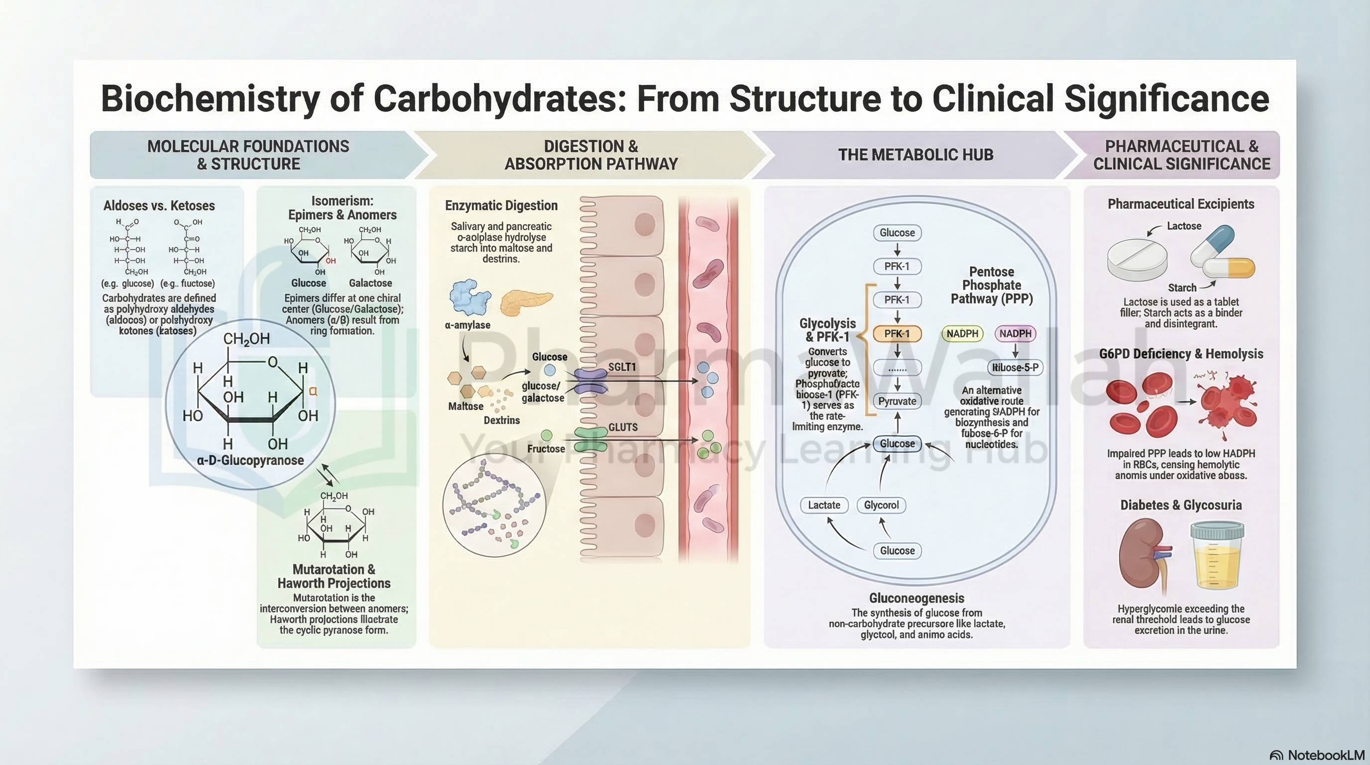
Definition: Carbohydrates are polyhydroxy aldehydes or ketones, or substances that yield such compounds upon hydrolysis. They have the general formula Cₙ(H₂O)ₙ (hydrates of carbon).
Chemical Nature:
- Contain carbon, hydrogen, and oxygen.
- Functional groups: Aldehyde (-CHO) or Ketone (-C=O) groups with multiple hydroxyl (-OH) groups.
- The simplest carbohydrates are monosaccharides (e.g., glucose, fructose).
2.2 Classification of Carbohydrates
| Class | Subtype | Description | Examples |
|---|---|---|---|
| Monosaccharides | Aldoses | Contain aldehyde group | Glucose, Galactose, Mannose |
| Ketoses | Contain ketone group | Fructose, Ribulose | |
| Oligosaccharides | Disaccharides | 2 monosaccharide units | Sucrose, Maltose, Lactose |
| Trisaccharides | 3 monosaccharide units | Raffinose | |
| Polysaccharides | Homopolysaccharides | Same repeating units | Starch, Glycogen, Cellulose |
| Heteropolysaccharides | Different repeating units | Hyaluronic acid, Heparin |
Based on Carbon Chain Length:
- Trioses (3C): Glyceraldehyde, Dihydroxyacetone
- Tetroses (4C): Erythrose
- Pentoses (5C): Ribose, Deoxyribose
- Hexoses (6C): Glucose, Fructose, Galactose
- Heptoses (7C): Sedoheptulose
2.3 Optical Activity of Carbohydrates
- Definition: The ability of a compound to rotate plane-polarized light.
- Chiral Centers: Carbohydrates contain asymmetric carbon atoms (chiral centers) that give rise to optical isomers.
- D and L Isomers: Based on the configuration of the asymmetric carbon farthest from the carbonyl group:
- D-isomer: OH group on the right (most naturally occurring sugars).
- L-isomer: OH group on the left.
- Enantiomers: Mirror-image isomers (e.g., D-glucose and L-glucose).
- Diastereomers: Non-mirror image isomers (e.g., D-glucose and D-mannose).
- Epimers: Differ at only one chiral carbon (e.g., D-glucose and D-galactose at C-4).
- Anomers: Cyclic forms differing at the anomeric carbon (C-1 for aldoses):
- α-anomer: OH at C-1 is below the ring plane.
- β-anomer: OH at C-1 is above the ring plane.
- Mutarotation: The change in optical rotation observed when an α or β anomer is dissolved in water and reaches equilibrium (a mixture of both anomers).
2.4 Important Reactions of Carbohydrates
| Reaction Type | Description | Example/Application |
|---|---|---|
| Oxidation | Aldehyde group oxidized to carboxylic acid | Glucose → Gluconic acid (mild oxidizing agents) |
| Strong oxidation | Both ends oxidized | Glucose → Glucaric acid |
| Reduction | Carbonyl group reduced to alcohol | Glucose → Sorbitol (used in sugar-free products) |
| Esterification | Hydroxyl groups react with acids | Formation of phosphate esters (e.g., glucose-6-phosphate) |
| Glycosidic Bond Formation | Anomeric carbon reacts with alcohol | Formation of disaccharides and polysaccharides |
| Osazone Formation | Reaction with phenylhydrazine | Characteristic crystalline derivatives for identification |
| Fermentation | Anaerobic breakdown by enzymes | Glucose → Ethanol + CO₂ (yeast) |
| Molisch Test | General test for carbohydrates | Purple ring at interface (positive for all carbs) |
| Benedict's Test | Reducing sugars | Brick-red precipitate (glucose, fructose, lactose) |
2.5 Biological Importance of Carbohydrates
- Primary Energy Source: Glucose is the main fuel for cellular respiration (ATP production).
- Energy Storage: Glycogen (in animals) and starch (in plants) store glucose.
- Structural Role: Cellulose (plants), Chitin (exoskeleton), and Peptidoglycan (bacterial cell walls).
- Cell Recognition: Glycoproteins and glycolipids on cell surfaces mediate cell-cell interactions and immune responses.
- Nucleic Acid Backbone: Ribose and deoxyribose form the structural framework of RNA and DNA.
- Metabolic Intermediates: Important in the synthesis of other biomolecules.
2.6 Pharmaceutical Importance of Carbohydrates
| Pharmaceutical Application | Example |
|---|---|
| Excipients and Fillers | Lactose, starch, dextrose used in tablet formulations |
| Drug Delivery Systems | Chitosan and hyaluronic acid for targeted delivery |
| Diagnostic Agents | Glucose tolerance test for diabetes mellitus |
| Antibiotics | Aminoglycosides (e.g., streptomycin) contain sugar moieties |
| Vaccine Development | Polysaccharide-based vaccines (e.g., Pneumococcal vaccine) |
| Anticoagulants | Heparin (a heteropolysaccharide) prevents blood clotting |
| Suspending Agents | Acacia, tragacanth in liquid formulations |
| Dietary Supplements | Glucose, fructose used in parenteral nutrition |
PART B: LIPIDS
2.7 Chemistry & Definition
Definition: Lipids are a heterogeneous group of organic compounds that are relatively insoluble in water (hydrophobic) but soluble in non-polar organic solvents like chloroform, ether, and benzene.
Chemical Nature:
- Primarily composed of carbon, hydrogen, and oxygen, with occasional phosphorus and nitrogen.
- They are esters of fatty acids with alcohols (e.g., glycerol, cholesterol).
2.8 Classification of Lipids
A. Based on Saponification (Hydrolysis by Alkali)
| Type | Definition | Examples |
|---|---|---|
| Saponifiable Lipids | Contain ester bonds that can be hydrolyzed by alkali to yield fatty acids and alcohols. | Triglycerides, Phospholipids, Sphingolipids, Waxes |
| Non-Saponifiable Lipids | Do not contain ester bonds and cannot be hydrolyzed by alkali. | Cholesterol, Steroids, Terpenes, Fat-soluble vitamins (A, D, E, K) |
B. Based on Chemical Composition
| Class | Subtype | Components | Examples |
|---|---|---|---|
| Simple Lipids | Fats & Oils (Triglycerides) | Glycerol + 3 Fatty acids (esters) | Animal fats, vegetable oils |
| Waxes | Fatty acid + Long-chain alcohol | Beeswax, Lanolin | |
| Complex Lipids | Phospholipids | Glycerol + 2 Fatty acids + Phosphate + Nitrogen base | Lecithin, Cephalin |
| Glycolipids | Glycerol/Sphingosine + Fatty acid + Carbohydrate | Cerebrosides, Gangliosides | |
| Lipoproteins | Lipid + Protein | HDL, LDL, VLDL | |
| Derived Lipids | Steroids, Terpenes | Hydrocarbon rings and side chains | Cholesterol, Bile acids, Vitamin D, Steroid hormones |
2.9 Chemistry of Fatty Acids
Definition: Fatty acids are carboxylic acids with long hydrocarbon chains (usually 4–36 carbons). They are the building blocks of many lipids.
Classification of Fatty Acids:
| Basis | Type | Description | Examples |
|---|---|---|---|
| Chain Length | Short-chain (SCFA) | 4–10 carbons | Butyric acid (4:0) |
| Medium-chain (MCFA) | 10–14 carbons | Lauric acid (12:0) | |
| Long-chain (LCFA) | 14–24 carbons | Palmitic acid (16:0), Stearic acid (18:0) | |
| Degree of Saturation | Saturated Fatty Acids (SFA) | No double bonds (single bonds only) | Palmitic acid, Stearic acid |
| Unsaturated Fatty Acids (USFA) | Contain one or more double bonds | Oleic acid, Linoleic acid | |
| Monounsaturated (MUFA) | One double bond | Oleic acid (18:1) | |
| Polyunsaturated (PUFA) | ≥2 double bonds | Linoleic acid (18:2), Arachidonic acid (20:4) | |
| Essential vs Non-essential | Essential Fatty Acids (EFA) | Cannot be synthesized by the body; must be obtained from diet | α-Linolenic acid (Omega-3), Linoleic acid (Omega-6) |
| Non-essential Fatty Acids | Can be synthesized by the body | Palmitic acid, Stearic acid | |
| Configuration | Cis-fatty acids | H atoms on same side of double bond | Naturally occurring (e.g., Oleic acid) |
| Trans-fatty acids | H atoms on opposite sides of double bond | Industrially produced (hydrogenation) |
Essential Fatty Acids (EFA):
- α-Linolenic acid (ALA) – Omega-3 family (18:3)
- Linoleic acid (LA) – Omega-6 family (18:2)
- Arachidonic acid – Conditionally essential (20:4) (can be synthesized from LA)
- Eicosapentaenoic acid (EPA) and Docosahexaenoic acid (DHA) – Long-chain Omega-3s (found in fish oil)
2.10 Important Reactions of Lipids and Fatty Acids
| Reaction Type | Description | Significance |
|---|---|---|
| Saponification | Alkaline hydrolysis of ester bonds | Formation of soap (salts of fatty acids) and glycerol |
| Hydrogenation | Addition of H₂ across double bonds (unsaturated → saturated) | Solidification of vegetable oils (margarine production) |
| Halogenation | Addition of halogens at double bonds | Used to determine the degree of unsaturation (Iodine number) |
| Hydrolysis | Breakdown by water (enzymatic) | Digestion of dietary fats by lipases |
| Oxidation (Rancidity) | Auto-oxidation of unsaturated fatty acids | Development of unpleasant odor and taste (spoilage) |
| Esterification | Reaction with alcohols to form esters | Formation of triglycerides, phospholipids |
| Acrolein Test | Dehydration of glycerol | Test for glycerol-containing lipids |
| Liebermann-Burchard Test | Reaction with acetic anhydride and sulfuric acid | Test for cholesterol (green color) |
2.11 Biological Importance of Lipids
- Energy Source: Most concentrated source of energy (9 kcal/g vs. 4 kcal/g for carbohydrates).
- Energy Storage: Fats stored in adipose tissue are a major reserve fuel.
- Structural Role: Phospholipids and cholesterol form the lipid bilayer of cell membranes.
- Insulation: Subcutaneous fat provides thermal insulation and mechanical cushioning.
- Hormone Precursors: Cholesterol is the precursor for steroid hormones (estrogen, testosterone, cortisol).
- Bile Acid Synthesis: Cholesterol forms bile acids necessary for fat digestion and absorption.
- Fat-soluble Vitamin Carriers: Vitamins A, D, E, K are lipid-soluble and absorbed with dietary fats.
- Eicosanoid Synthesis: Essential fatty acids are precursors to prostaglandins, thromboxanes, and leukotrienes (mediators of inflammation and immunity).
2.12 Pharmaceutical Importance of Lipids
| Pharmaceutical Application | Example |
|---|---|
| Drug Formulation | Oils and fats used as vehicles for hydrophobic drugs in ointments, creams, and emulsions |
| Liposomal Drug Delivery | Liposomes (phospholipid vesicles) used for targeted drug delivery |
| Parenteral Nutrition | Lipid emulsions (Intralipid) provide essential fatty acids in IV nutrition |
| Cholesterol-Lowering Drugs | Statins (HMG-CoA reductase inhibitors) target cholesterol biosynthesis |
| Bile Acid Sequestrants | Drugs like cholestyramine bind bile acids to lower cholesterol |
| Diagnostic Agents | Lipid profile tests measure total cholesterol, HDL, LDL, triglycerides for cardiovascular risk assessment |
| Drug Absorption Enhancement | Lipid-based formulations improve bioavailability of poorly soluble drugs |
| Emulsifying Agents | Lecithin used as an emulsifier in pharmaceuticals and cosmetics |
PART C: PROTEINS AND AMINO ACIDS
2.13 Chemistry & Definition
Definition: Proteins are large, complex macromolecules composed of amino acids linked by peptide bonds. They are the most abundant organic molecules in the human body, constituting about 50% of cellular dry weight.
Chemical Nature:
- Contain carbon, hydrogen, oxygen, nitrogen, and often sulfur (in cysteine and methionine).
- Protein molecules are polymers of amino acids joined by peptide bonds (-CO-NH-).
- The diversity of proteins arises from the sequence and arrangement of 20 standard amino acids.
2.14 Classification of Amino Acids
A. Based on R-group Properties (Chemical Nature)
| Class | Characteristics | Amino Acids |
|---|---|---|
| Non-polar (Hydrophobic) | Aliphatic or aromatic R-groups; insoluble in water | Glycine (Gly), Alanine (Ala), Valine (Val), Leucine (Leu), Isoleucine (Ile), Methionine (Met), Proline (Pro), Phenylalanine (Phe), Tryptophan (Trp) |
| Polar (Hydrophilic) | Neutral but have polar groups; soluble in water | Serine (Ser), Threonine (Thr), Asparagine (Asn), Glutamine (Gln), Tyrosine (Tyr), Cysteine (Cys) |
| Acidic (Negatively Charged) | Side chain contains carboxyl group | Aspartic acid (Asp), Glutamic acid (Glu) |
| Basic (Positively Charged) | Side chain contains amino group | Lysine (Lys), Arginine (Arg), Histidine (His) |
B. Based on Nutritional Requirements (Biosynthetic Capacity)
| Type | Definition | Amino Acids |
|---|---|---|
| Essential | Cannot be synthesized by the body; must be obtained from diet | Valine, Leucine, Isoleucine, Methionine, Phenylalanine, Threonine, Tryptophan, Lysine, Histidine* (*essential in children) |
| Non-Essential | Can be synthesized by the body | Glycine, Alanine, Serine, Aspartic acid, Glutamic acid, Asparagine, Glutamine, Proline, Cysteine, Tyrosine |
| Conditionally Essential | Essential in certain disease states or in children | Arginine, Histidine, Tyrosine, Cysteine |
C. Based on Metabolic Fate (Ketogenic vs Glucogenic)
| Type | Fate | Amino Acids |
|---|---|---|
| Glucogenic | Can be converted to glucose (via gluconeogenesis) | Alanine, Glycine, Serine, Aspartate, Glutamate (most amino acids) |
| Ketogenic | Can be converted to ketone bodies/fatty acids | Leucine, Lysine |
| Both (Glucogenic & Ketogenic) | Can be converted to both glucose and ketones | Isoleucine, Phenylalanine, Tryptophan, Threonine, Tyrosine |
2.15 Classification of Proteins
A. Based on Shape and Solubility
| Type | Description | Examples |
|---|---|---|
| Fibrous Proteins (Scleroproteins) | Long, thread-like molecules; insoluble in water; structural role | Collagen (bone, tendons), Keratin (hair, nails, skin), Elastin (ligaments), Myosin (muscle) |
| Globular Proteins (Albumins) | Spherical, compact molecules; water-soluble; functional role | Hemoglobin (oxygen transport), Immunoglobulins (immune function), Enzymes, Albumin (osmotic balance) |
B. Based on Chemical Composition
| Type | Definition | Examples |
|---|---|---|
| Simple Proteins | Composed only of amino acids | Albumin, Globulins, Histones, Protamines |
| Conjugated Proteins | Contain amino acids + a non-protein prosthetic group | Glycoproteins (protein + carbohydrate), Lipoproteins (protein + lipid), Hemoproteins (protein + heme), Metalloproteins (protein + metal ion) |
| Derived Proteins | Degradation products of simple/conjugated proteins | Proteoses, Peptones, Peptides |
2.16 Organizational Levels of Protein Structure
| Level | Description | Bonds/Forces Involved | Example |
|---|---|---|---|
| Primary Structure | Linear sequence of amino acids in a polypeptide chain | Peptide bonds | Insulin: A-chain (21 aa), B-chain (30 aa) |
| Secondary Structure | Folding of the polypeptide chain into local regular structures | Hydrogen bonds (between backbone groups) | α-helix (hair-like spiral), β-pleated sheet (zigzag) |
| Tertiary Structure | Overall three-dimensional shape of a single polypeptide chain | Hydrophobic interactions, Hydrogen bonds, Disulfide bonds, Ionic bonds, Van der Waals forces | Myoglobin, Immunoglobulins |
| Quaternary Structure | Spatial arrangement of two or more polypeptide subunits | Same as tertiary (inter-subunit interactions) | Hemoglobin (α₂β₂ tetramer), DNA polymerase |
2.17 Macromolecular Nature of Proteins
- Molecular Weight: Proteins range from 5,000 Da (small peptides) to over 1,000,000 Da (large complexes).
- Polymerization: Composed of 50 to several thousand amino acid residues.
- Conformation: Proteins exist in native (functional) and denatured (non-functional) states.
- Biological Activity Depends on Conformation: The specific 3D structure determines function; loss of native conformation (denaturation) leads to loss of biological activity.
Denaturation of Proteins:
- Definition: Disruption of higher-order (secondary, tertiary, quaternary) structure without breaking peptide bonds.
- Agents: Heat, pH extremes, organic solvents, heavy metal ions, detergents.
- Examples: Egg white coagulation on heating; irreversible in most cases.
2.18 Important Reactions of Amino Acids and Proteins
| Reaction/Test | Reagent | Positive Result | Amino Acid/Group Detected |
|---|---|---|---|
| Ninhydrin Reaction | Ninhydrin | Purple/Blue color | All amino acids (α-amino group) |
| Biuret Test | Biuret reagent (Cu²⁺ in alkaline solution) | Violet color | Proteins and peptides (≥2 peptide bonds) |
| Millon's Reaction | Millon's reagent (Hg(NO₃)₂) | Red color | Tyrosine (phenolic group) |
| Xanthoproteic Reaction | Concentrated HNO₃ (heat) followed by NaOH | Yellow → Orange color | Aromatic amino acids (Phenylalanine, Tyrosine, Tryptophan) |
| Sakaguchi Reaction | α-Naphthol + Sodium hypochlorite | Red color | Arginine (guanidinium group) |
| Nitroprusside Test | Sodium nitroprusside | Purple/Violet color | Cysteine (sulfhydryl -SH group) |
| Hopkins-Cole Reaction | Glyoxylic acid + H₂SO₄ | Purple ring | Tryptophan (indole ring) |
| Ehrlich's Reaction | p-Dimethylaminobenzaldehyde (p-DMAB) | Blue color | Tryptophan (indole ring) |
| Sorenson's Formol Titration | Formaldehyde + NaOH | Quantification of amino acids | Amino groups (used for titration) |
2.19 Biological Importance of Proteins
- Enzymes: Catalyze virtually all biochemical reactions (e.g., DNA polymerase, digestive enzymes).
- Structural Support: Collagen (bone, skin), Keratin (hair, nails), Elastin (elastic tissues).
- Transport: Hemoglobin (O₂ transport), Albumin (fatty acid transport), Transferrin (iron transport).
- Immunological Defense: Immunoglobulins (antibodies) protect against pathogens.
- Hormones and Signaling: Insulin, Glucagon, Growth hormone, Cytokines.
- Movement and Contraction: Actin, Myosin (muscle contraction).
- Gene Regulation: Histones (DNA packaging), Transcription factors.
- Blood Clotting: Fibrinogen, Thrombin.
- Osmotic Balance: Albumin maintains oncotic pressure in blood.
- Buffering: Proteins (e.g., Hemoglobin) act as biological buffers.
2.20 Pharmaceutical Importance of Proteins and Amino Acids
| Pharmaceutical Application | Example |
|---|---|
| Therapeutic Proteins | Insulin (diabetes), Erythropoietin (anemia), Monoclonal antibodies (cancer, autoimmune disease), Interferons (viral infections) |
| Enzyme Replacement Therapy | Alpha-1 antitrypsin (emphysema), Laronidase (Mucopolysaccharidosis) |
| Vaccines | Protein-based vaccines (e.g., Hepatitis B surface antigen) |
| Drug Targets | Many drugs target proteins: Receptors, Enzymes (e.g., ACE inhibitors for hypertension), Ion channels |
| Amino Acid Supplements | Glutamine (gut health), Arginine (wound healing), Branched-chain amino acids (muscle recovery) |
| Diagnostic Markers | Serum albumin (liver/kidney disease), Myoglobin (muscle injury), Troponin (heart attack), HbA1c (diabetes monitoring) |
| Parenteral Nutrition | Amino acid infusions for patients unable to eat (TPN) |
| Drug Design | Understanding protein structure enables rational drug design (e.g., HIV protease inhibitors) |
| Gene Therapy | Proteins as delivery vectors or therapeutic gene products |
| Biopharmaceuticals | Insulin, growth hormones, clotting factors (recombinant DNA technology) |
4. Diagrams/Tables
Table 1: Summary of Carbohydrates Classification
                              CARBOHYDRATES
                                    │
            ┌───────────────────────┼───────────────────────┐
            │                       │                       │
     MONOSACCHARIDES          OLIGOSACCHARIDES       POLYSACCHARIDES
        (1 unit)                (2–10 units)            (>10 units)
            │                       │                       │
       - Aldoses              - Disaccharides      - Homopolysaccharides
       - Ketoses              - Trisaccharides     - Heteropolysaccharides
            │                       │                       │
        Glucose                  Sucrose                Starch
        Fructose                 Maltose                Glycogen
        Galactose                Lactose                Cellulose
        Ribose                                          Hyaluronic acid
| Classification | Subtypes | Examples |
|---|---|---|
| Monosaccharides (1 unit) | Aldoses, Ketoses | Glucose, Fructose, Galactose, Ribose |
| Oligosaccharides (2–10 units) | Disaccharides, Trisaccharides | Sucrose, Maltose, Lactose |
| Polysaccharides (>10 units) | Homopolysaccharides, Heteropolysaccharides | Starch, Glycogen, Cellulose, Hyaluronic acid |
Table 2: Protein Structure Hierarchy
| Level | Structure | Key Features | Stabilizing Forces |
|---|---|---|---|
| Primary | Linear chain of amino acids | Sequence determines all higher levels | Peptide bonds (covalent) |
| Secondary | Local folding (α-helix, β-sheet) | Regular repeating patterns | Hydrogen bonds (backbone) |
| Tertiary | 3D folding of one chain | Globular or fibrous shape | Hydrophobic, H-bonds, S-S, Ionic |
| Quaternary | Assembly of multiple chains | Subunit interactions | Same as tertiary |
Diagram 1: Structure of an Amino Acid
        H
        │
H₂N ─── C ─── COOH
        │
        R
R = Variable side chain
Key Groups
- Amino group:
-NH₂ - Carboxyl group:
-COOH - α-carbon: Central carbon atom
- R-group: Variable side chain that distinguishes each amino acid
Diagram 2: Peptide Bond Formation
      H₂N ── CH ── COOH   +   H₂N ── CH ── COOH
              │                       │
              R₁                      R₂

                    ↓ Condensation

      H₂N ── CH ── CO ── NH ── CH ── COOH   +   H₂O
              │               │
              R₁              R₂
Peptide bond: -CO-NH-
5. Exam Angle
Short Questions (2-5 marks)
- Classify carbohydrates with suitable examples.
- What is optical activity? Explain D and L configuration of sugars.
- Differentiate between saponifiable and non-saponifiable lipids.
- List essential fatty acids and their pharmaceutical importance.
- What is the primary structure of proteins?
- What is denaturation of proteins? Give causes and examples.
- Differentiate between glucogenic and ketogenic amino acids.
- Write the test for tyrosine and cysteine.
- What are conjugated proteins? Give examples.
- Differentiate between α-helix and β-pleated sheet.
Long Essays (10+ marks)
- Classify carbohydrates, describe their chemical reactions, and explain their biological and pharmaceutical importance.
- Write a detailed note on the classification of lipids and the pharmaceutical importance of lipids.
- Describe the organizational levels of protein structure. Add a note on the biological and pharmaceutical importance of proteins.
- Give the classification of amino acids and describe their chemical reactions.
Key Points Frequently Tested
- Carbohydrates: Reducing properties, mutarotation, D/L isomerism.
- Lipids: Essential fatty acids (Omega-3, Omega-6), saponification number, rancidity.
- Proteins: Names and abbreviations of 20 amino acids, denaturation, tests for specific amino acids.
- Clinical correlations: G6PD deficiency (carbohydrate metabolism), hyperlipidemia, protein-energy malnutrition, rheumatoid arthritis (protein biomarkers).
6. Summary Box
- Carbohydrates are polyhydroxy aldehydes/ketones classified as mono-, oligo-, and polysaccharides. They exhibit optical activity and provide energy, structure, and cell recognition.
- Lipids are hydrophobic molecules classified as simple, complex, or derived lipids. Essential fatty acids (Omega-3, Omega-6) must be obtained from the diet. Lipids serve as energy stores, membrane components, and hormone precursors.
- Proteins are macromolecules made of amino acids linked by peptide bonds. There are 20 standard amino acids, classified by R-group properties and nutritional requirements.
- Protein structure has four levels: primary (sequence), secondary (α-helix/β-sheet), tertiary (3D folding), and quaternary (subunit assembly). The native conformation is essential for function.
- All three classes of biomolecules have immense pharmaceutical importance—as drugs, excipients, diagnostic markers, drug targets, and therapeutic agents.
7. References
- Chatterjee, M. N., & Shinde, R. (2011). Textbook of Medical Biochemistry (8th ed.). Jaypee Brothers Medical Publishers.
- Murray, R. K., Bender, D. A., Botham, K. M., Kennelly, P. J., Rodwell, V. W., & Weil, P. A. (2015). Harper's Illustrated Biochemistry (30th ed.). Lange Medical Books / McGraw-Hill Education.
- Nelson, D. L., & Cox, M. M. (2012). Lehninger Principles of Biochemistry (6th ed.). W. H. Freeman.
- Peet, A., Lieberman, M. A., & Marks, A. (2012). Marks' Basic Medical Biochemistry (4th ed.). Lippincott Williams & Wilkins.
- Champe, P. C., & Harvey, R. A. (2013). Lippincott's Illustrated Reviews: Biochemistry (6th ed.). Lippincott Williams & Wilkins.
End of unit 2 of 5
Check what you've learned
5 quick questions from this unit. They're optional: try them if you like, then mark the unit as read and move on.